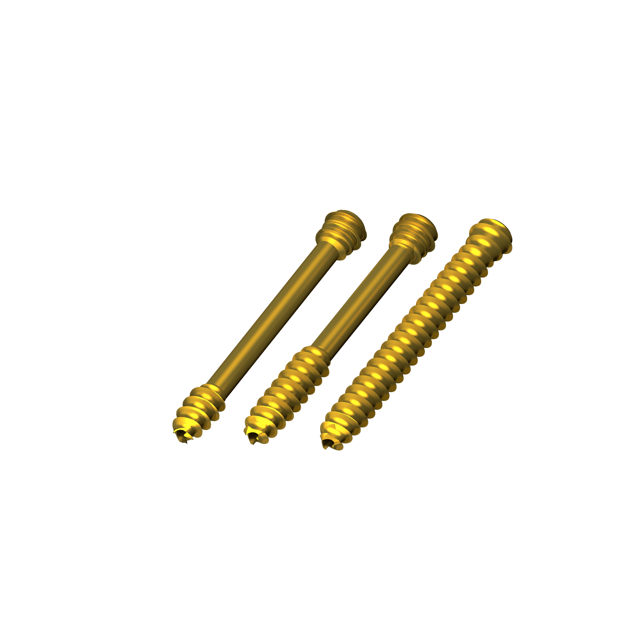
Designed to Perform
The APTUS Ankle Trauma System 2.8/3.5 is designed to provide all the low profile straight and anatomically pre-contoured plates for all surgical approaches, fractures and osteotomies around the ankle.
Anatomical Design

All anatomical pre-contoured plates are designed according to an extensive database to accommodate different patient anatomies.
Anatomical Flaps

Flaps in the Lateral Fibula and Anterolateral Tibia plates allow for capture of the Wagstaffe-LeFort fragment and Tillaux-Chaput tubercle.
Specialised Instrumentation

Controlled reduction of the syndesmosis.
TriLock

Patented TriLock locking technology provides multidirectional locking of the screw in the plate.
Approaches
The APTUS Ankle Trauma System 2.8/3.5 is designed for plate insertion in all the surgical approaches around the ankle including specific MIPO instrumentation.
TriLock Distal Fibula Plates 2.8/3.5, Lateral

Design Highlights
-
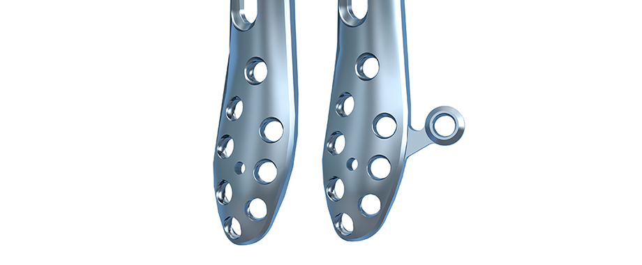
Plate is available with and without flap for Wagstaff fixation.
-
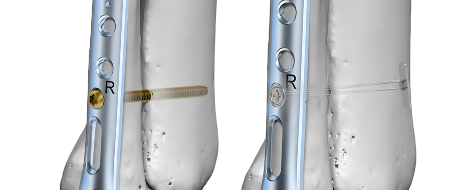
20° posterior-anterior oblong hole and single syndesmotic hole for placement of syndesmotic screws or suture button implants (single hole).
-
Plate position can be adjusted on the bone before final fixation.
-
Bone plates can be temporarily held in position during screw fixation of the plate.
-
1.6 mm thick plates with offset screw geometry in the shaft.
-
Eight distal screw fixation options for fixation of comminuted fractures or extra fixation in osteoporotic bone.
TriLock Distal Fibula Plates 2.8, Straight

Design Highlights
-
The oblong hole allows for the plate position to be adjusted before final fixation.
-
Plates are 1.6 mm thick with an offset screw geometry.
-
Standard plate has three screw placements in the distal section of the plate when extra fixation is needed.
TriLock 3.5, Straight

Design Highlights
-
Fibula fractures with syndesmosis injuries can be stabilized using the 2-, 3- and 4-hole 3.5 TriLock straight plates.
-
Holes are designed to fit the lateral button of a syndesmosis suture implant, allowing the button to sit flush within the plate.
TriLock Distal Tibia Plates 2.8/3.5, Medial

Design Highlights
-
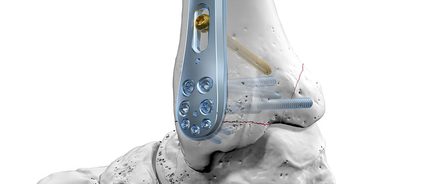
Head of plate has multiple 2.8 mm and 3.5 TriLock and Cortical screws for the capture of small and large bone fragments.
-
Thin distal metaphyseal section accepts 2.8 mm screws for malleolar fracture fixation.
-
Plate can also be used for SMO with 2 - 3 mm axial compression through the compression hole.
TriLock Distal Tibia Plates 2.8/3.5, Anterolateral

Design Highlights
-
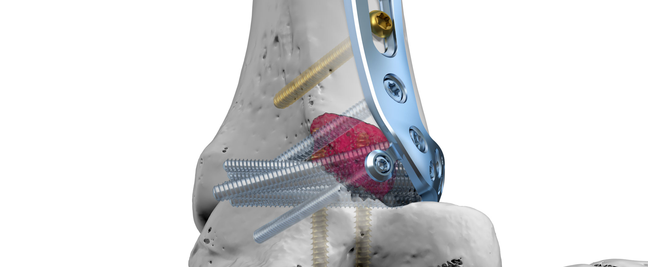
2.8 flap for capture of Chaput fragment if required.
-
Double row of screws in the distal head accept 2.8 mm and 3.5 TriLock and Cortical screws for rafting of the tibial plafond.
TriLock Distal Tibia Plates 3.5, T and L

Design Highlights
-
Distal holes are angled 10° proximally to prevent penetration of the joint.
-
Reconstruction plate design allows for the plates to be bent 3D.
Technological Milestones in Osteosynthesis
TriLock
Multidirectional and angular stable locking, screws can pivot freely by ± 15° in all directions
TriLockPLUS
TriLockPLUS screw holes offer the advantage of locking and compression in one step
HexaDrive
Simplified screw pick-up due to self-holding system
Storage

Customized system arrangement and modular concept.
- Easy to use and sterilize
- Lightweight components
- Validated cleaning and sterilization of the implants
Explore Various Perspectives
APTUS Ankle – Surgical Technique Animation
APTUS Ankle – Posterolateral Approach
APTUS Ankle – Medial Approach
APTUS Ankle - Anterior Approach
APTUS Ankle – Lateral Malleolar Approach
Documentation
Results
- Ankle Trauma System 2.8 / 3.5 – Surgical Technique 22.01.2024 10 MB Surgical Technique Lower Extremities English
- Case Study – Ankle Fracture, Fibula Plate 2.8/ 3.5 Tibia T-Plate, Dr. Joshua A. Sebag, US 17.11.2023 1 MB Case Study Lower Extremities English
- Case Study – Ankle Fracture, Fibula Plate 2.8, Dr. Joshua A. Sebag, US 17.11.2023 879 KB Case Study Lower Extremities English
- Case Study – Ankle Fracture, 2.8 Fibula Plate & 3.5 Tibia T-Plate, Dr. Tim Schepers, Netherlands 17.11.2023 161 KB Case Study Lower Extremities English
- Case Study – STUDYFixation of a Supination External Rotation IV Trimalleolar Ankle Fracture 17.11.2023 1 MB Case Study Lower Extremities English
- APTUS Ordering Catalog/Bestellkatalog/Catalogue (High Resolution) (Version 0, MDR) 18.01.2024 22 MB Ordering Catalog Upper Extremities English
- APTUS Ordering Catalog/Bestellkatalog/Catalogue (Low Resolution) (Version 0, MDR) 18.01.2024 16 MB Ordering Catalog Upper Extremities English
- Ankle Trauma System 2.8, 3.5 – Product Information 17.11.2023 674 KB Product Information Lower Extremities English
- Ankle Trauma 2.8, 3.5 – White Paper 17.11.2023 407 KB White paper Lower Extremities English


