Precision and Innovation
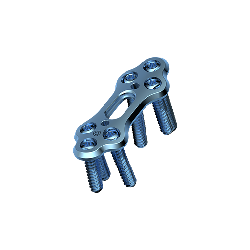
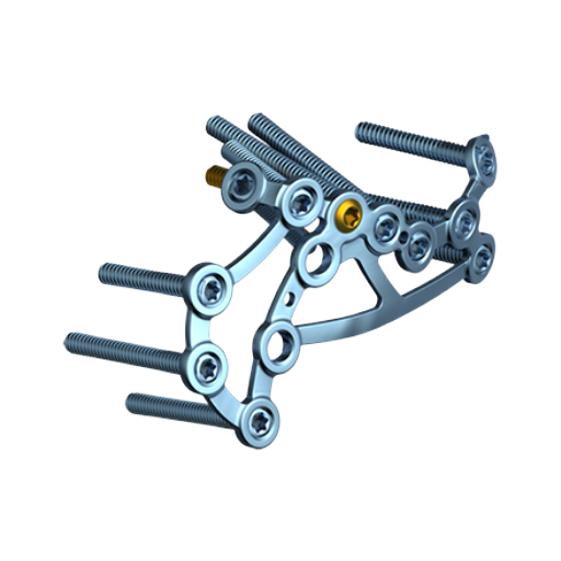
The APTUS Hallux system was developed in association with a team of internationally renowned foot surgeons. The plate designs were confirmed using statistical analysis of CT datasets and were thoroughly verified on anatomical specimens. Covering TMT-1 arthrodesis via dorsomedial and plantar fixation with dedicated plates for classic or modified Lapidus. As well as plates for MTP-1 arthrodesis; plates for primary approach and revision cases enables surgeons to face complex cases.
Anatomical Fit

Plate shape derived from the results of extensive anatomical analyses.
Plate Design

Made up from a titanium block. Sophisticated production techniques enable Medartis manufacturing complex anatomical shapes without bending process.
TriLock Plus

Compression and angular stable locking in one step.
TriLock

TriLock locking technology provides multidirectional and angular stable locking of the screw in the plate.
Treatment Concept

TriLock TMT-1 Medial Fusion Plates 2.8

Highlights
-
Available in two sizes with a plate thickness of 1.6 mm and 2.0 mm
-
Plate design reduces contact with the tibialis anterior tendon
-
Versatile approach, classical or modified Lapidus technique in the same plate due to the 4.0 transfixation screw
-
Compression by means: (1) TriLockPlus (2) K-wire slot for compression with Olive K-wires
-
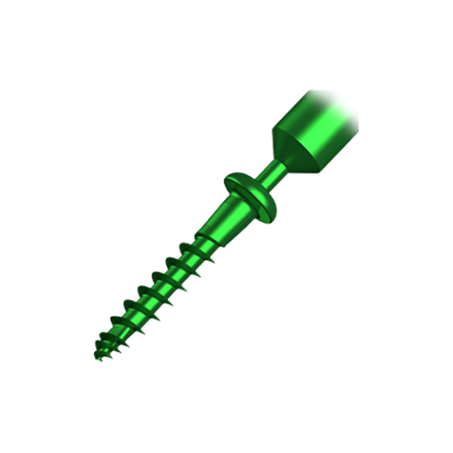
4.0 Transfixation screw with SpeedTip technology compatible with 2.8 instruments, as the SpeedTip tip design widens the drilled hole
Features
TriLock TMT-1 Plantar Fusion Plates 2.8

Highlights
-
Plate positioning minimizes overlapping with the insertion of the tibital anterior tendon*
-
Anatomical plate shape makes bending almost unnecessary*
-
Screw alignment along with the TriLock multidirectionality allow soft tissue friendly access
* (Plaass et al.: "Placement of Plantar Plates for Lapidus Arthrodesis Anatomical Considerations" Seven plantar plate designs were analyzed on 29 anatomic specimens with regard to plate position towards the tibialis anterior tendon, general plate design as well as the necessity of further bending. The APTUS plantar fusion plate achieved the best result.
Features
TriLock Grid Plates 2.8

Highlights
-
Generic form and therefore the plate can be adapted for a wide range of indications
-
Double row of screws in the plate-head area which increases subchondral stability
Features

TriLock MTP-1 Fusion Plates 2.8

Highlights
-
Three defined dorsiflexion angles (0°, 5°, 10°) with 10º of valgus inclination
-
Compression by means: (1) TriLockPlus (2) K-wire slot for compression with Olive K-wires
-
Reduced likelihood of collisions of transverse screws
-
Additional proximal plate hole for increased primary stability in poor bone quality
-
Crossing lag or CCS screw can be placed if needed
Features
TriLock MTP-1 Revision Plates 2.8

Highlights
-
Two defined dorsiflexion angles (5°, 10°) with 10º of valgus inclination
-
Compression by means: (1) TriLockPlus (2) K-wire slot for compression with Olive K-wires
-
Closely arranged distal holes enable treatment even of small fragments
-
Oblong hole allows for variable autograft fixation
Features
Technological Milestones in Osteosynthesis
TriLock
Multidirectional and angular stable locking, screws can pivot freely by ± 15° in all directions
TriLockPLUS
TriLockPLUS screw holes offer the advantage of locking and compression in one step
HexaDrive
Simplified screw pick-up due to self-holding system
Designed to Organize

- Compact
- Completely modular
- Compact system
- Easy to use
- Clear storage overview of implants and instruments
- Validated cleaning and sterilization
Explore Various Perspectives
MTP Revision Procedure with the 2.8 TriLock MTP Revision Plate
Medartis Patientenstory MTP Fusion Peter Neuenschwander
Hallux valgus correction with TMT-1 instability
MTP Fusion with two crossing CCS 4.0 Screws
Fusion of the MTP 1 Joint
MTP Revision Procedure with the 2.8 TriLock MTP Revision Plate
Documentation
Results
- Hallux System 2.8 – Surgical Technique 22.01.2024 5 MB Surgical Technique Lower Extremities English
- APTUS Ordering Catalog/Bestellkatalog/Catalogue (High Resolution) (Version 0, MDR) 18.01.2024 22 MB Ordering Catalog Upper Extremities English
- APTUS Ordering Catalog/Bestellkatalog/Catalogue (Low Resolution) (Version 0, MDR) 18.01.2024 16 MB Ordering Catalog Upper Extremities English
- Hallux System 2.8 – Product Information (incl. Ordering Information) 17.11.2023 6 MB Product Information Lower Extremities English


